

By the women’s health team at Renu IVF, Kanpur | Medically reviewed by Dr. Renu Singh Gahlaut, IVF
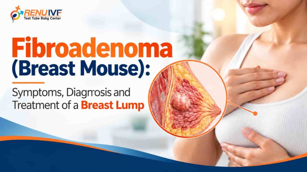
Finding a lump in your breast is frightening. Many women quietly worry for weeks before telling anyone, because the first thought is cancer. The truth is more reassuring: most breast lumps are benign, and one of the most common causes, especially in younger women, is a fibroadenoma. You may also hear it called a breast mouse, because it feels small, smooth and slippery, and slides away under the fingers.
This guide explains what a breast fibroadenoma is, how it feels, how it differs from other causes of a breast lump, how doctors confirm the diagnosis, which treatment options exist, what changes in pregnancy and fertility treatment, and which warning signs should send you to a doctor without delay. It is written for readers in India, so it also addresses the common “breast me ganth” worry directly and in simple Hinglish. All medical points are drawn from Mayo Clinic, the American Cancer Society, MedlinePlus, Ireland’s HSE and a published systematic review, and rewritten here in our own words.
| Quick answerA fibroadenoma is a solid, non-cancerous breast lump that is usually firm, smooth, rubbery, round and easy to move under the skin. It is most common between ages 15 and 35 and is often painless. Many need no treatment, only monitoring. But a lump cannot be judged by touch alone: every new breast lump should be examined by a doctor and, usually, confirmed with an ultrasound or mammogram and sometimes a biopsy. |
| Watch first: Breast Mouse explained by Dr. Renu Singh GahlautBreast Mouse क्या होता है? | Fibroadenoma के लक्षण, जाँच और इलाज | Dr. Renu Singh GahlautShort video from the Dr Renu Singh Gahlaut YouTube channel explaining what a breast mouse (fibroadenoma) is, how to recognise its symptoms, how it is investigated and how it is treated, and why a new lump should always be checked.Watch the video on YouTube |
What Is a Fibroadenoma? Understanding the Breast Fibroadenoma
A fibroadenoma is a benign (non-cancerous) growth made of two things the name describes: glandular tissue (adeno) and fibrous connective tissue (fibro). It forms inside the breast and is not an infection, not an injury and not a sign that something you did caused it. Doctors do not know the exact cause, but hormones that control the menstrual cycle are thought to play a role, which fits with how often fibroadenomas appear in teenagers and young women (Mayo Clinic).
How common is it? MedlinePlus describes fibroadenoma as the commonest benign breast tumour and the commonest breast tumour in women under 30. The American Cancer Society notes that they are most often found in women in their 20s and 30s but can occur at any age, and Mayo Clinic gives the usual range as 15 to 35 years.
Why is it called a “breast mouse”?
“Breast mouse” is a popular, informal name. A typical fibroadenoma is smooth and mobile, so when you press on it, it can slide away under the skin like a tiny mouse. This is the feeling many women describe. The name is useful for recognising the feel, but it should never be used to decide that a lump is harmless: other lumps, including some that need treatment, can also feel mobile.
Breast Lump: Not Every Lump Is a Fibroadenoma
A breast lump can have many causes. HSE says most lumps are harmless, such as a fibroadenoma or a fluid-filled cyst, but some can be breast cancer, and it advises against trying to work out the cause yourself. Mayo Clinic also lists fibrocystic breast changes, a noncancerous tumour called a hamartoma, injury and mastitis among benign causes. The table below shows how different causes tend to behave. It is a guide to understanding, not a way to diagnose yourself.
| Possible cause | Typical feel and clues | Needs checking? |
| Fibroadenoma | Firm, smooth, rubbery, round or oval, well-defined edges, moves easily; usually painless; most often ages 15 to 35 | Yes, to confirm the diagnosis |
| Breast cyst | Fluid-filled sac; smooth, may be tender; can change with the menstrual cycle | Yes, ultrasound shows whether it is fluid or solid |
| Fibrocystic changes | General lumpiness or thickening, often tender and cyclical | Yes, to rule out a separate lump |
| Infection or mastitis | Red, warm, painful area, sometimes with fever, common in breastfeeding | Yes, same day if fever or severe pain |
| Injury or fat necrosis | Lump after a knock or surgery; may be firm or bruised | Yes, because it can resemble other lumps on scans |
| Phyllodes tumour | Rare; firm lump that grows fairly quickly and may stretch the skin; most common in the 40s | Yes, promptly; may need removal for diagnosis |
| Breast cancer | May be hard, irregular or fixed, but can also feel smooth; may come with skin dimpling, nipple change or bloody discharge | Yes, urgently |
The key point is in the last column: the answer is “yes” every time. Touch cannot tell a cyst from a fibroadenoma from a cancer, and even experienced doctors need imaging and sometimes a biopsy.
Fibroadenoma Symptoms: What Does It Feel Like?
Many fibroadenomas are found by accident, during a shower, while dressing, or on a scan done for another reason. Based typical fibroadenoma has these features:
• Shape and feel: round or oval, firm and rubbery, with smooth, well-defined edges. Some women compare it to a pea, a marble or a flat coin.
• Mobility: it moves easily under the skin when you press on it.
• Pain: usually painless, although it may feel tender in the days before a period.
• Size: about 2.5 cm (1 inch) on average, though some are too small to feel and others reach several centimetres.
• Number and side: most women have one, but some have several, in one or both breasts.
• Behaviour over time: it often stays the same or grows slowly. It may enlarge in pregnancy and often shrinks after menopause.
| A reminder about lumps that feel “typical”A lump that fits this description is likely to be a fibroadenoma, but “likely” is not “confirmed”. The same article that describes the typical feel also tells readers to see a provider for any new lump. Confirmation needs a clinical examination and tests. |
Breast Fibroadenoma Types: Simple, Complex, Juvenile and Giant
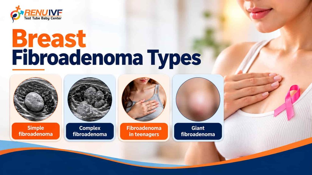
Doctors describe several patterns, mostly based on how the lump looks under the microscope or how it behaves.
| Type | What it means | Why it matters |
| Simple fibroadenoma | The most common type; looks uniform under the microscope | Does not appear to raise breast cancer risk (American Cancer Society) |
| Complex fibroadenoma | Shows extra “proliferative” changes; tends to be larger and more often seen in older women | May raise risk very slightly; screening advice generally unchanged. Can grow and press on nearby tissue (Mayo Clinic) |
| Fibroadenoma in teenagers | Usually found around puberty; often called a juvenile fibroadenoma | Many shrink or disappear over months to a few years (Mayo Clinic), so doctors often watch carefully |
| Giant fibroadenoma | Grows quickly and can be larger than about 5 cm (2 inches) | May distort the breast; often needs evaluation for removal |
| Phyllodes tumour (separate condition) | Rare tumour of the breast’s supporting tissue; can resemble a fibroadenoma on scans | Most are benign, but some are borderline or malignant; needs complete removal in many cases |
Can a fibroadenoma turn into cancer?
This is the question most women are afraid to ask. The reassuring answer is that common (simple) fibroadenomas do not appear to increase the risk of breast cancer. Complex fibroadenomas and phyllodes tumours may raise it slightly, according to Mayo Clinic, and the American Cancer Society says there are no specific risk figures it can give for complex lesions. Two practical points follow. First, because a fibroadenoma can look similar to a phyllodes tumour on a scan, a biopsy or removal is sometimes needed to be sure. Second, the lump you find may turn out, rarely, not to be a fibroadenoma at all. Both are reasons to get the diagnosis confirmed rather than assumed.
Causes and Risk Factors
No one knows exactly why fibroadenomas form. What the sources agree on is the connection with hormones:
• They are most common in girls going through puberty and in women of reproductive age, and they are much less common after menopause (MedlinePlus).
• They may grow in pregnancy and tend to shrink after menopause, which points to hormonal influence (American Cancer Society).
• MedlinePlus adds that a fibroadenoma may not shrink after menopause in women who take hormone therapy.
• They are not caused by something you ate, a bra, a knock, or by lifting heavy weights. No such link is described in the medical sources used for this article.
This matters emotionally. Many women blame themselves, or are told by relatives that they brought it on through diet or habits. A fibroadenoma is not a reflection of your lifestyle.
Breast Me Ganth: Kya Har Ganth Cancer Hoti Hai?
Short jawab: nahi. Zyadatar breast ki ganth benign hoti hai, jaise fibroadenoma ya cyst. Lekin kuch ganth cancer ho sakti hain, aur sirf haath se chhoone par ye farq pata nahi chalta. Isliye “kuch nahi hoga” ya “cancer hi hoga”, dono soch galat hain. Sahi tareeka hai jaldi doctor ko dikhana.
Bharat mein aksar mahilayen sharm, dar, ya gharelu dabav ki wajah se ganth ko mahino tak chhupaye rakhti hain. Kuch log massage, jadi-booti ya bina jaanch ki dawai se theek karne ki koshish karte hain. Isse sirf waqt kharab hota hai. HSE ke anusaar, ganth mehsoos hote hi use check karwana chahiye, kyunki cancer jitni jaldi pakda jaye, utna aasan ilaaj hota hai.
Kab turant doctor ko dikhana chahiye?
• Nayi ganth, ya purani ganth jo badh rahi ho ya kathor hoti ja rahi ho.
• Ganth jo agle periods ke baad bhi na jaye (Mayo Clinic).
• Breast ka size ya shape badalna, ya skin mein dimple ya santre ke chhilke jaisi skin.
• Nipple andar ki taraf dhans jana, ya nipple se khoon ya saaf paani nikalna.
• Breast ya nipple par lalima, rash ya crusting.
• Armpit mein ganth ya sujan.
Agar report mein fibroadenoma aaya hai, tab bhi follow-up aur doctor ki salah zaroori hai. Aur agar aap IVF, IUI ya pregnancy ki planning kar rahi hain, to apni ganth ke baare mein fertility doctor ko pehle bata dein.
How a Fibroadenoma Is Diagnosed: The Three-Step Check
HSE describes the standard approach used at breast clinics: a breast examination, a scan (usually ultrasound or mammogram), and a needle biopsy where needed. Doctors often call this a “triple assessment”. The three parts support one another, and together they are far more reliable than any single test.
| Step | What happens | What it tells doctors |
| 1. Clinical breast examination | The doctor asks about the lump, your cycle, pregnancy, family history and medicines, then examines both breasts and the armpits | Size, shape, mobility, firmness, skin or nipple changes and lymph nodes |
| 2. Imaging | Ultrasound, mammogram or both. In younger women ultrasound is usually the first scan | Whether the lump is solid or fluid-filled, its borders and shape, and whether anything else is present |
| 3. Needle biopsy | A small sample of tissue is taken with a needle, guided by ultrasound or mammogram, after numbing the area | The definite tissue diagnosis: fibroadenoma, phyllodes tumour, cyst-related change or cancer |
Do teenagers and young women always need a biopsy?
Not always. MedlinePlus notes that teenagers and women in their early 20s may skip the biopsy if the lump disappears or remains unchanged over time. Your doctor makes this call from the examination and ultrasound findings.
Why biopsy is sometimes not enough
The American Cancer Society explains that phyllodes tumours can be hard to separate from fibroadenomas on imaging, and that a core biopsy sometimes cannot settle the question, so the whole lump may need to be removed for testing. A systematic review in Breast Care lists features that make doctors suspect a phyllodes tumour rather than a fibroadenoma: a persistent lump, pain, rapid growth, a dense mass on mammography, or round clefts or cysts on ultrasound.
How to read your report
Radiology reports often include a category score called BI-RADS. In general, lower numbers mean the lump looks benign and higher numbers mean it needs more attention, and the report ends with a recommendation such as routine follow-up, a repeat scan after a few months, or a biopsy. Ask the doctor who ordered the scan to explain your score and the plan in plain language.
Fibroadenoma Treatment: Watch, Remove or Ablate?
Treatment is individual. The American Cancer Society says not all fibroadenomas need it, and MedlinePlus explains that removal is optional in many cases. The table summarises how doctors usually decide.
| Approach | Usually considered when | What to know |
| Monitoring (watchful waiting) | Small, stable lump with no symptoms and no worrying features on imaging or biopsy | Repeat clinical exams and scans at intervals your doctor sets. One review mentions follow-up over roughly 6 to 12 months and notes that attendance for follow-up is often poor, so keep your appointments |
| Surgical removal (excision) | Large lump, growth over time, pain or other symptoms, distortion of the breast, uncertain or atypical findings, or a concern about a phyllodes tumour | Done under anaesthesia. Risks include scarring, change in the nipple area, loss of breast volume and anaesthetic risks (Breast Care review) |
| Vacuum-assisted removal | Selected small lumps that are clearly benign | Minimally invasive and described as safe and effective; suitability depends on the lump and the centre |
| Cryoablation or radiofrequency ablation | Selected small lumps confirmed as fibroadenomas | Destroys the lump with cold or heat, guided by ultrasound, without removing it (MedlinePlus; American Cancer Society) |
| High-intensity focused ultrasound (HIFU) | Still emerging; used in some centres | A systematic review reported a technical success rate of about 93%, with lump volume falling by roughly 35% to 62% at 6 months, and called for larger, longer studies |
Is there a medicine or home remedy that dissolves a fibroadenoma?
The medical sources used for this article list monitoring, surgical removal and ablation as the options. None of them lists tablets, creams, massage or herbal products as a treatment. If someone advises you to “dissolve it” with such products instead of getting scans, treat that advice with caution, because it delays the diagnosis that actually protects you.
After treatment: what to expect
• Keep follow-up appointments. If part of a lump is left behind, or new tissue forms, it should be checked.
• New fibroadenomas can develop after one is removed. The American Cancer Society stresses that this does not mean the old lump returned, but any new lump should still be examined.
• Phyllodes tumours, even benign ones, can come back in the same place, so closer follow-up with exams and scans is usual.
Fibroadenoma, Pregnancy, Breastfeeding and Fertility Treatment
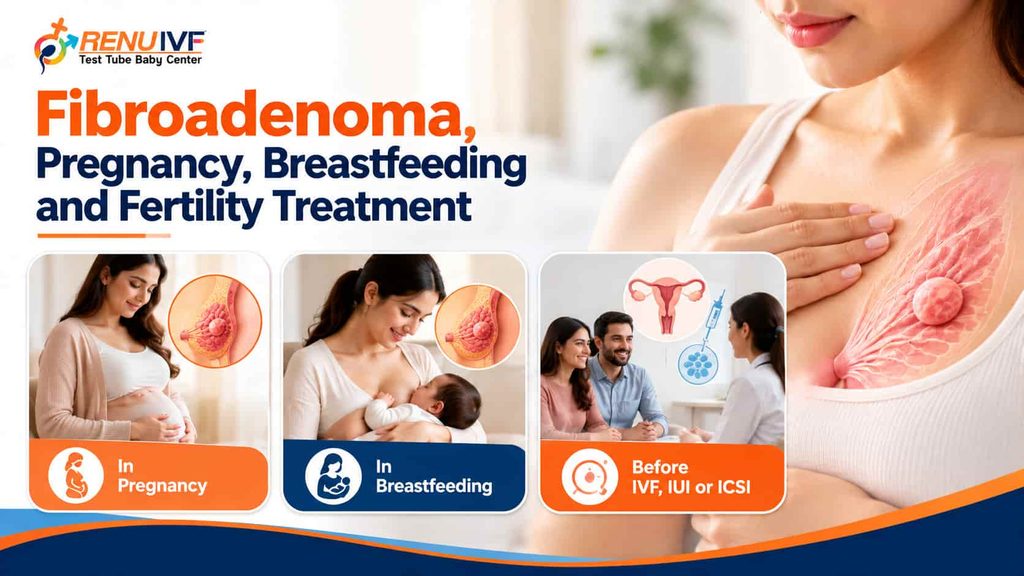
Because fibroadenomas are influenced by hormones, women often ask what happens when pregnancy or fertility treatment begins.
In pregnancy and breastfeeding
Mayo Clinic and the American Cancer Society both note that fibroadenomas can enlarge during pregnancy. Most such changes are benign, but pregnancy also changes breast tissue, so a lump that is new, hard or growing quickly should be examined rather than assumed to be an old fibroadenoma. Ultrasound uses no radiation and is usually the first scan. Your doctor decides on any further tests, and treatment, if needed, is planned around your pregnancy or feeding schedule.
Before IVF, IUI or ICSI
A fibroadenoma is not known to be a cause of infertility. However, fertility treatment can raise the body’s estrogen levels, and hormones influence breast tissue, so many fertility specialists prefer that any new lump is examined before treatment starts. There is no strong evidence that fertility medicines cause fibroadenomas, but it is sensible to include a breast examination in your pre-treatment review. A structured fertility assessment is a good point to raise this, and our patient-education articles on the IVF journey explain what to expect at each stage. If you are planning IUI, IVF or ICSI, tell your doctor about any lump, past biopsy or family history of breast disease.
In a high-risk pregnancy
If a breast lump is found while you are pregnant, or if you already have other pregnancy risks, ask your obstetrician whether your care plan needs a breast specialist’s input. Our guidance on high-risk pregnancy care and our Pregnancy Care and Complications library cover this wider picture.
Red Flags: When a Breast Lump Needs Urgent Attention
MedlinePlus agree on the changes that should prompt a prompt visit. Seeing a doctor does not mean something is wrong. It means a professional can check it early.
| Urgency | Signs | What to do |
| See a doctor as soon as possible | • A new lump or thickening in the breast or armpit• A known lump that grows, firms up or changes shape• A lump that does not go after your next period• Change in breast size or shape• Skin dimpling, puckering or an orange-peel texture• A nipple that turns inward when it did not before• Clear or bloody nipple discharge• Redness, rash or crusting on the nipple or breast skin• Unexplained bruising of the breast | Book an examination within days. Do not wait for the next period, a “second opinion” at home or another month of observation. |
| Same day | • Hot, red, painful breast with fever• A lump that is very painful or rapidly enlarging• A lump with an open sore or bleeding | Seek urgent care or call your doctor the same day. |
Breast Awareness and Screening
The aim is not to perform a perfect self-test every month. It is to know what is normal for your breasts so that you notice a change quickly. HSE recommends getting to know how your breasts look and feel at different times in your cycle. Mayo Clinic Health System adds that self-exams are less effective than routine mammograms at detecting cancer or improving survival, so awareness complements screening and does not replace it.
A simple way to check
1. Choose a consistent time. For women who menstruate, a few days after the period ends is when breasts are least tender and lumpy.
2. Look first. Stand in front of a mirror with arms down, then raised. Look for changes in size, shape, skin dimpling, redness or nipple position.
3. Feel with the pads of your fingers. Use light, medium and firm pressure, moving in small circles or straight lines across the whole breast, from the collarbone to the bra line and into the armpit.
4. Note what you find. A new lump, a change in an existing lump, or anything that feels different from the other side should be written down and shown to a doctor.
5. Do not squeeze the nipple. Report any spontaneous discharge instead.
Mammograms and age
Mayo Clinic Health System says women at average risk are advised yearly screening mammograms starting at age 40, with earlier or more frequent screening for women with a family history or other risk factors. Guidelines differ between countries and organisations, so ask your doctor what is right for you. Screening is for women without symptoms. A lump needs a diagnostic work-up, not routine screening.
Myths and Facts
| Myth (aam galatfehmi) | Fact |
| “Breast me ganth ka matlab cancer hai.” | Most breast lumps are benign, but you cannot tell by touch. Every new lump needs a check. |
| “Fibroadenoma hai to operation karna hi padega.” | No. Small, stable lumps are often just monitored. Removal is for large, growing, symptomatic or uncertain lumps. |
| “Massage ya dawai se ganth ghul jayegi.” | Reliable sources list monitoring, removal and ablation. None list massage or tablets as a treatment. |
| “Ek baar nikal gayi to dobara nahi hogi.” | New fibroadenomas can form later. A new lump does not mean the old one came back, but it still needs checking. |
| “Ye galat khaan-paan ya bra ki wajah se hoti hai.” | The cause is unknown, hormones are thought to play a role, and no such lifestyle link is described in the sources used. |
| “Fibroadenoma hai to pregnancy nahi ho sakti.” | A fibroadenoma is a benign breast lump and is not known to cause infertility. Tell your doctor, especially before fertility treatment. |
| “Agar ganth dard nahi karti to koi problem nahi.” | Many fibroadenomas and many cancers are painless. Pain is not a reliable guide. |
| “Ultrasound mein theek aaya to biopsy ki zarurat nahi.” | Sometimes a biopsy is still needed, because imaging cannot always separate a fibroadenoma from a phyllodes tumour. |
Living With a Fibroadenoma: Practical Tips
• Keep a record. Note the date you found the lump, its size on the scan report, and any change. Keep copies of every report.
• Attend every follow-up. A watched lump is only safe if it is actually watched.
• For cyclical tenderness, a well-fitting supportive bra and a warm compress can ease discomfort. Ask your doctor before using any pain medicine, especially if you are pregnant or trying to conceive.
• Do not poke or squeeze the lump repeatedly. It can cause pain and make it harder to judge changes.
• Tell every doctor about your fibroadenoma, including your gynaecologist, fertility specialist and obstetrician.
• Look after your general health. Balanced nutrition, regular activity and enough sleep support overall wellbeing, as discussed in our nutrition and lifestyle articles.
How to Prepare for Your Appointment
A little preparation helps the doctor make faster and better decisions.
• Write down when you first noticed the lump, how it has changed, and whether it changes with your period.
• Note any pain, nipple discharge, skin change or armpit swelling.
• List all medicines, hormonal pills, injections and supplements, and whether you are pregnant, breastfeeding or planning treatment for fertility.
• Note any family history of breast, ovarian or other cancers.
• Bring earlier scan or biopsy reports and, if possible, the images themselves.
• Wear a comfortable two-piece outfit so that examination is easy.
Questions worth asking
• What do you think this lump is, and what do we need to confirm it?
• Which tests do I need, and when will I get the results?
• If it is a fibroadenoma, does it need treatment or only follow-up?
• How often should I be re-examined or re-scanned?
• Does this affect my pregnancy plans, IVF or breastfeeding?
• Which signs should make me return sooner?
A simple lump diary
| Date | Location and size (approx.) | Pain or tenderness? | Change since last note | Cycle day / pregnancy |
Frequently Asked Questions About Fibroadenoma and Breast Lumps
What is a fibroadenoma?
A fibroadenoma is a solid, non-cancerous breast lump made of glandular and connective tissue. It is typically firm, smooth, rubbery and easy to move under the skin, and it is usually painless. Mayo Clinic says it most often appears between ages 15 and 35, and MedlinePlus calls it the most common benign breast tumour.
Why is a fibroadenoma called a “breast mouse”?
“Breast mouse” is an informal name. A fibroadenoma usually slips easily under the fingers when you press it, as if it were a small mouse running away. It is not a medical term, and doctors will use the word fibroadenoma in your reports.
Breast me ganth ka matlab kya hai, aur kya har ganth cancer hoti hai?
Breast me ganth ka matlab hai breast mein koi lump ya gaanth mehsoos hona. Har ganth cancer nahi hoti. Zyadatar lumps benign hote hain, jaise fibroadenoma ya cyst, lekin kuch ganth cancer ho sakti hain, aur sirf haath se chhoone par farq pata nahi chalta. Isliye har nayi ganth ko doctor se check karwana chahiye.
Can a fibroadenoma turn into cancer?
Ordinary (simple) fibroadenomas do not appear to raise breast cancer risk. The American Cancer Society says complex fibroadenomas may raise risk slightly, but screening advice generally stays the same. Phyllodes tumours, which can look like fibroadenomas on scans, are a separate group in which some are cancerous, which is why a biopsy matters.
Does a fibroadenoma go away on its own?
Sometimes. Mayo Clinic notes that many fibroadenomas in teenagers shrink over months to a few years, and fibroadenomas often shrink after menopause. In other people the lump stays the same size or grows slowly, so doctors follow it up.
Does every fibroadenoma need surgery?
No. MedlinePlus explains that a small, stable lump that causes no symptoms and does not look suspicious can be left in place and monitored. Removal is considered if the biopsy is unclear, the lump is painful or growing, or cancer is a concern.
At what size should a fibroadenoma be removed?
There is no single size cut-off. Doctors weigh the size, growth, symptoms, your age, imaging and biopsy findings. For context, Mayo Clinic describes giant fibroadenomas as lumps that grow quickly and can pass about 5 cm (2 inches), and the American Cancer Society lists large size, growth, distortion of the breast and symptoms among reasons to consider treatment.
Can a fibroadenoma grow during pregnancy?
Yes, it can. Both Mayo Clinic and the American Cancer Society note that fibroadenomas can enlarge during pregnancy, and they are believed to respond to hormones. Any new or fast-growing lump in pregnancy or while breastfeeding should be examined rather than watched at home.
Is a fibroadenoma painful?
Usually it is painless, although some are tender, especially in the days before a period. Persistent pain is one of the reasons a doctor may consider removing the lump.
Can a fibroadenoma come back after removal?
The same lump is not expected to return after complete removal, but new fibroadenomas can form later. The American Cancer Society points out that a new lump does not mean the old one regrew, so every new lump still needs to be checked.
What is the difference between a fibroadenoma and a breast cyst?
A fibroadenoma is a solid growth of breast tissue, while a cyst is a fluid-filled sac. They can feel similar to the touch, which is why an ultrasound is usually the first scan: it can show whether a lump is solid or fluid-filled.
What is the difference between a fibroadenoma and a phyllodes tumour?
Phyllodes tumours are rare tumours that start in the supporting (stromal) tissue of the breast. According to the American Cancer Society, they are hard to tell apart from fibroadenomas on imaging and often grow faster. Most are benign, but some are borderline or malignant, so a rapidly growing lump should be assessed promptly.
Can a fibroadenoma affect fertility, IVF or breastfeeding?
A fibroadenoma is a benign breast lump and is not known to be a cause of infertility. Because hormones can influence breast tissue, it is sensible to have any new lump examined before fertility treatment, pregnancy or breastfeeding, and to tell your doctor if you already know you have one.
Which doctor should I see for a breast lump?
Start with a doctor who can examine you and arrange imaging: a gynaecologist, a general or breast surgeon, or a breast clinic. Your doctor may refer you for ultrasound, mammogram or a biopsy. At Renu IVF in Kanpur you can book a consultation for women’s health concerns and be guided on the next step.
Can I treat a fibroadenoma at home with medicines or massage?
Reliable medical sources describe monitoring, surgical removal and image-guided ablation as the options. They do not list tablets, creams or massage as a treatment, and pressing or massaging a lump can cause pain and confusion about whether it has changed. Get it checked first, and follow your doctor’s advice.
Next Steps and Care at Renu IVF, Kanpur
A breast lump does not have to be a source of fear. Most are benign, many need only follow-up, and the ones that need treatment are best handled early. The single most useful step is to have it examined.
At Renu IVF in Kalyanpur, Kanpur, Dr. Renu Singh Gahlaut and the care team see women for fertility, pregnancy and gynaecological concerns. If you have found a breast lump, especially if you are planning pregnancy or fertility treatment, book a consultation and we will guide you on the examination and the right specialist or imaging centre for your situation. You can also watch more patient-education videos in our video gallery and read more in our Gynecological Health and Fertility Issues and Conditions articles. We welcome patients from Unnao, Kanpur Dehat, Fatehpur, Kannauj, Etawah and Hardoi.
| Watch again and shareBreast Mouse क्या होता है? | Fibroadenoma के लक्षण, जाँच और इलाज | Dr. Renu Singh GahlautWatch the video on YouTube |